
At VetTriage we assist pets and their owners worldwide. For our clinic partners and patients in Australia, snakes bites can be an all too common occurance. The issue is, diagnosing a snake bites is quite tricky. In order to confirm the attack actually occurred, the pet owner would have to visualize the snake interaction with their pet. Otherwise treatment could be delayed and a misdiagnosis could result. Additionally, the human pet owner is also at risk for a snake bite, so awareness is vital. The following will review the current Australian veterinary medical literature as well as elaborations for many of the terms used.
Overall incidence
In reported Australian human-animal bite incidences, a wide variety of animals are implicated. Snakes (42% of human bites), dogs (29%), and cats (9%) are the most common bites to humans.1 Therefore as a pet owner, one must be aware of their surroundings at all times, for themselves and for their pets.
In Australia, there is an estimated 6,200 dog-snake bite cases reported annually.2 Out of 28 hospitals from 6 Australian states/territories, 624 cases were entered into a registry, including 419 dogs (67% of cases) and 205 cats (33% of cases) involved with snake bites.3 It is no surprise that the incidence of snake bites to dogs is greater than that of cats; dogs tend to be more curious and friendly, and they tend to explore the outdoors with the pet owner at a greater frequency than is true with cats.
Overall, the approximate rate of dog-snake bites is 11.4 dogs per year in Australia.4 Within New South Wales, Australia the yearly prevalence of snake envenomation in dogs is estimated at 0.31%.5 In Australia, approximately 84% of cases occurs in the 6 warmer months of the year.4 Bites are more prominent in rural (78% incidence) than urban areas (22% incidence).2

Most dogs are of the sporting breed.4 Retrievers, spaniels and pointers are such canine breed examples (see image above). These breeds are bred for hunting and a highly active lifestyle. The average age of dogs affected is 3.6 years.4 Younger dogs, generally speaking, tend to be more active outdoors and tend to be more curious and inexperienced, lending themselves to more trouble. Bites occur in the owner’s yard in 85% of dog cases and in 26% of cat cases.3 In dog and cat cases, 42% of pets are reported to be bit in the 3 hours between 03:00 PM and 05:59 PM.3
Snake venom and snake species
Venom consists of 90% water and has a minimum of 10 enzymes and 3 to 12 nonenzymatic proteins and peptides in any individual snake.6 At least 50 enzymes may contribute to snake venom potency. These enzymes cause soft tissue damage, vasculotoxicity, coagulopathy, cytotoxicity, and necrosis.7 Soft tissue damage refers to that of the skin, muscles and related tissues where the snake bite wound occur. Vascular or blood vessel damage released to a toxin describes vasculotoxicity. A coagulopathy is a general term to describe any abnormality or derangements with the body’s ability to form a clot and stop bleeding from continuing. Any damage to cells by a toxin or chemical is termed cytotoxic. Necrosis is cellular death due to any pathology.
The elapids are a family of venomous snakes characterized by their permanently erect fangs at the front of the mouth.15 So for example, the elapid venom may be divided into the components of prothrombin activating enzymes, lipases and peptidic neurotoxins.5
The action of these venom components may result in neurotoxic (pre-synaptic and post-synaptic), hemotoxic (red-cell destruction and coagulation disturbance), cardiovascular, myotoxic (muscle toxicity) and secondary nephrotoxic (kidney toxicity) effects.5
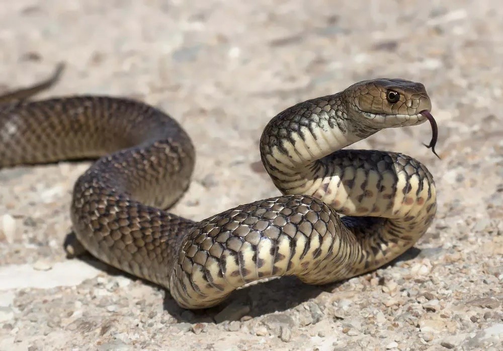
In Australia, the eastern brown snake (Pseudonaja textilis, see the image at the top of the article) is responsible for an estimated 76% of reported snakebite cases to domestic pets nationally each year.2,11 In another report, the brown snake is implicated in 3.7% of cases.4

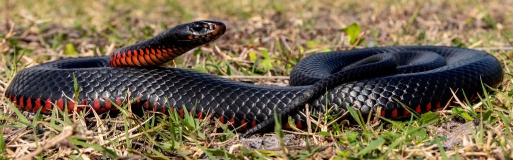
Tiger snakes (Notechis scutatus pictured above) are implicated in 13 to 40% of cases in Australia.2,4 Black snakes (Pseudechis porphyriacus, see below) account for 6% of cases.2
The wide variation in incidences may be due to the discrepancies in reporting, the absolute or correct identification of a snake/specific snake species and the specifics of the region where the data was collected.
In New South Wales, Australia the most common species reported to be responsible for envenomation is the red-bellied black snake followed by the brown snake and then the tiger snake.5
History and clinical signs
The onset of clinical signs after envenomation may be delayed for several hours. The presence of fang marks does not indicate that envenomation has occurred, only that a bite has taken place.6 In Australia, approximately 64% of dogs are actually seen to be bit or in contact with a snake.4 The median time from the moment the bite occurs to presentation at the veterinary hospital is 60 minutes in dogs and 95 minutes in cats.3
The most common presenting clinical signs are ptyalism (salivation), emesis (vomiting), mydriasis (dilated pupils), absence of the pupillary light reflex, depression and generalized muscle weakness, hindlimb ataxia (neurologic incoordination) and respiratory distress.4 Tissue swelling at the bite site occurs in 53% of cases and facial palsy is seen in 12% of cases.8 The median elapsed time from hospital admission to onset of hemoptysis (coughing blood) is 2 hours (range 0 to 18 hours).9
With cases involving black snakes, the prominent gross findings (meaning those abnormalities noted on a physical examination) include icterus (a yellow discoloration to the skin and mucous membranes due to red blood cell destruction and/or liver/gall bladder disease), localized facial edema (swelling due to tissue fluid accumulation as a marker for inflammation) in the region of the presumed bite wound, pigmenturia (urine discoloration) and multicavitary serosanguineous effusions (fluid accumulation within the body).10 With the eastern brown snake in south eastern Queensland, Australia, pulmonary hemorrhage (bleeding in the lungs) can be seen based on clinical examination and overt hemoptysis.9
Diagnostics
Hematological profiles post-envenomation reveal anemia (low red blood cell count indicating bleeding, lack of red blood cell production, or red blood cell destruction) and spherocytosis (abnormality spherical red blood cells) in 100% of cases.8
With the eastern brown snake, the primary pathology is a venom-induced consumptive coagulopathy.9,11. Clinicopathological changes due to a coagulopathy may include hemolysis, increased creatine kinase (a muscle enzyme seen in elevated concentrations with muscle trauma or destruction), pigmenturia and mildly prolonged active clotting time (a blood test that evaluates the body’s ability to form clots).8
With the eastern brown snake), thoracic radiographs demonstrate a diffuse alveolar pattern, a nonspecific change that can be seen with any condition that causes inflammation of the lungs.9
Snake venom detection kits are used in only 1% to 28% of such cases.2,3
Histopathology (tissue examination) of black snake cases reveal acute renal tubular necrosis (kidney disease) with hemosiderosis (iron buildup from the destruction of red blood cells), marked splenic hemosiderosis and centrilobular to midzonal hepatocellular necrosis (liver disease) with severe cholestasis (gall bladder disease).10
Treatment
The most common treatment reported is a combination of intravenous fluid therapy (to treat dehydration), antivenom (the specific antidote or antiserum containing antibodies for that specific venom for that specific snake), and supportive care.4
Antivenom is also known as antivenene and antivenin. Antivenom is administered in 85% of cases and not used in 33% of cases.2,3 This may be due to cost, access to the product, or lack of knowledge that antivenom exists or that the patient’s clinical signs were from envenomation.
With the eastern brown snake envenomation, 80% of cases were managed with endotracheal intubation, 20% with mask oxygen supplementation alone, and 40% with mechanical ventilation.9 In dog snake bites overall, mechanical ventilation may be required in 11% of patients.8 These are various measures to treat dogs with severe respiratory compromise.
Fresh frozen canine plasma was administered to 70% affected by Eastern brown snake envenomation.9 Plasma is used to treat pets with coagulopathies among other medical indications. Whole blood transfusion may be required in 12% of patients to treat or combat anemia.8
Treatment response is based on the monitoring the patient’s response to treatment via the assessment of clinical signs.4 As such, every patient will have their own unique treatment depending on the details of the case and how their case progresses while being administered the treatments. The median hospitalization duration is 25 hours).3
Antivenin complications
Acute (meaning sudden or short-term) and chronic (over time or long-term) reactions are reported in 7% and 0.9% of dog cases, respectively. Death possibly due to these reactions occurs in 4.1% of dogs.12
Acute adverse reaction clinical signs may include facial swelling (4% incidence), sudden respiratory distress (1.9%) and vomiting (1.9%). The adverse reactions are self-limiting (meaning they resolve on their own without intervention or treatment) in 5.6% of the dogs, while 1.9% of dogs are treated with corticosteroids (glucocorticoids or anti-inflammatory steroids). No delayed adverse reactions believed to be associated with the antivenin administration are noted.14
Serum sickness is characterized by recurrent signs of a delayed hypersensitivity — swelling, edema, urticaria/hives, gastrointestinal signs and vasculitis (inflammation of blood vessels) one to two weeks after the antivenin has been administered. Treatment of serum sickness is the same as for any allergic reaction — glucocorticoids and antihistamines. Diagnosis is based on the history of receiving the antivenin, absence of other causes of allergic reaction, complement testing and skin biopsies to confirm vasculitis.13
Survival
An overall recovery rate of 87 to 88% is seen.3,9 With eastern brown snake envenomation, of the total number of cases presenting for treatment, 30% survive to hospital discharge, 60% are euthanized due to a poor prognosis and 10% die from cardiac arrest.9 In another study, 6% of dogs were euthanized and 94% survived to hospital discharge.8
Prognostic factors
The amount of time that passes between the bite and presenting to the veterinary hospital matters when it comes to the speed of recovery. For example, the period of time from treatment to full recovery is shorter for cases in which the time from the snake bite to beginning the treatment was one hour or less (24 hour recovery time in these dogs) when compared with the recovery time for all cases (36 hours recovery time in these dogs).4 Survival is also perceived to be associated with the time between envenomation and presentation to the veterinary clinic and with antivenom administration.5 This makes sense as the faster treatment can be instituted and the faster the antivenom can be administered, the faster the recovery.
The severity of clinical signs also affects prognosis. For example, the prognosis is poor for dogs presenting with the triad of complete flaccid paralysis, dyspnea (breathing difficulty) and a sub-normal temperature.4 With those dogs severely affected by respiratory compromise and requiring intense respiratory support, only 25% of these dogs survive to hospital discharge.9 Of those that require fresh frozen plasma treatment, 43% survive.9
The survival is also affected by the snake species involved. Survival after antivenom administration was reported to be highest for red-bellied black snakes.5
The treatment type also affects survival. Ninety-one percent of cats and 75% of dogs survive following the administration of antivenom, whereas only 66% of cats and 31% of dogs survive without the antivenom treatment.2 As a side note, cats apparently are more resistant to envenomation than dogs. While only 31% of dogs survive brown snake bites without antivenom, cats are twice as likely to survive bites (66%). Even with antivenom treatment, cats have a significantly higher survival rate.11
Conclusion
Snakebites are obviously a serious concern to humans, dogs, and cats. Chances are that if you reside in an area of the world where snakes, especially venomous snakes, our common place, the veterinary hospitals in that region are more likely to be equipped with the appropriate treatment, including anti-venom.
While in route to a veterinary hospital, it may be worthwhile calling them ahead of time to check on whether or not their staff is able to handle a potential snakebite case and whether or not the appropriate treatment is available.
Even more so, if you live in a high-risk area, plan ahead by knowing exactly which emergency veterinary hospital is equipped with anti-venom and experience with snakebites, so if this were to occur with your dog or cat, you know exactly where to go.
References
1. Vardanega J, Smith LK, Smith S, Hanson J. Animal bite wounds and their management in tropical Australia. Int J Infect Dis. 2022 Feb 18;118:1-9. doi: 10.1016/j.ijid.2022.02.026. Epub ahead of print. PMID: 35189338.
2. Mirtschin PJ, Masci P, Paton DC, Kuchel T. Snake bites recorded by veterinary practices in Australia. Aust Vet J. 1998 Mar;76(3):195-8. doi: 10.1111/j.1751-0813.1998.tb10128.x. PMID: 9578756.
3. Boller M, Kelers K, Stevenson MA, Winkel KD, Hardjo S, Heller J, Judge PR, Ong HM, Padula AM, Reddrop C, Santos L, Sharp CR, Smart L, Swindells KL, Tabrett D, Wierenga JR. SnakeMap: four years of experience with a national small animal snake envenomation registry. Aust Vet J. 2020 Sep;98(9):442-448. doi: 10.1111/avj.12993. Epub 2020 Aug 2. PMID: 32743816.
4. Hill FW. Snake bite in dogs. Aust Vet J. 1979 Feb;55(2):82-5. doi: 10.1111/j.1751-0813.1979.tb15171.x. PMID: 444166.
5. Heller J, Bosward KL, Hodgson JL, Cole FL, Reid SW, Hodgson DR, Mellor DJ. Snake envenomation in dogs in New South Wales. Aust Vet J. 2005 May;83(5):286-92. doi: 10.1111/j.1751-0813.2005.tb12743.x. PMID: 15957391.
6. Peterson ME. Snake bite: pit vipers. Clin Tech Small Anim Pract. 2006 Nov;21(4):174-82. doi: 10.1053/j.ctsap.2006.10.008. PMID: 17265901.
7. Armentano RA, Schaer M. Overview and controversies in the medical management of pit viper envenomation in the dog. J Vet Emerg Crit Care (San Antonio). 2011 Oct;21(5):461-70. doi: 10.1111/j.1476-4431.2011.00677.x. PMID: 22316194.
8. Finney ER, Padula AM, Leister EM. Red-bellied black snake (Pseudechis porphyriacus) envenomation in 17 dogs: clinical signs, coagulation changes, haematological abnormalities, venom antigen levels and outcomes following treatment with a tiger-brown snake antivenom. Aust Vet J. 2020 Jul;98(7):319-325. doi: 10.1111/avj.12953. Epub 2020 May 10. PMID: 32390184.
9. Leong OS, Padula AM, Leister E. Severe acute pulmonary haemorrhage and haemoptysis in ten dogs following eastern brown snake (Pseudonaja textilis) envenomation: Clinical signs, treatment and outcomes. Toxicon. 2018 Aug;150:188-194. doi: 10.1016/j.toxicon.2018.05.020. Epub 2018 May 29. PMID: 29857087
10. Kelly-Bosma M, Leister E, Padula A, Schaffer-White A, Bielefeldt-Ohmann H, Haworth M, Henning J, Allavena R. Pathology of Fatal Australian Black Snake (Pseudechis sp) Envenomation in Two Adult Dogs. J Comp Pathol. 2021 Jul;186:1-6. doi: 10.1016/j.jcpa.2021.04.009. Epub 2021 May 31. PMID: 34340798.
11. Zdenek CN, Llinas J, Dobson J, Allen L, Dunstan N, Sousa LF, Moura da Silva AM, Fry BG. Pets in peril: The relative susceptibility of cats and dogs to procoagulant snake venoms. Comp Biochem Physiol C Toxicol Pharmacol. 2020 Oct;236:108769. doi: 10.1016/j.cbpc.2020.108769. Epub 2020 May 3. PMID: 32376497.
12. McCown JL, Cooke KL, Hanel RM, Jones GL, Hill RC. Effect of antivenin dose on outcome from crotalid envenomation: 218 dogs (1988-2006). J Vet Emerg Crit Care (San Antonio). 2009 Dec;19(6):603-10. doi: 10.1111/j.1476-4431.2009.00487.x. PMID: 20017766.
13. Benjamin M Lee, Kristin M Zersen, Jennifer R Schissler, Lauren A Sullivan. Antivenin-associated serum sickness in a dog. J Vet Emerg Crit Care (San Antonio). 2019 Sep;29(5):558-563.
14. Lund HS, Kristiansen V, Eggertsdóttir AV, Skancke E, Ranheim B. Adverse reactions to equine-derived F(ab’)2 -antivenin in 54 dogs envenomated by Vipera berus berus. J Vet Emerg Crit Care (San Antonio). 2013 Sep-Oct;23(5):532-7. doi: 10.1111/vec.12099. Epub 2013 Sep 19. PMID: 24102941.
15. Elapidae. https://en.wikipedia.org/wiki/Elapidae. April 12, 202.


